吴玮莹,学号SY1410210,系我院生物医学工程专业生物力学与力生物学课题组14级硕士研究生,于2015年4月在期刊《Computerized Medical Imaging and Graphics》上发表题为《A two-layer structure prediction framework for microscopy cell detection》高水平学术论文一篇,该期刊影响因子为1.534。
课题背景及意义:
细胞显微结构和外形的识别在生物学和临床上都有重要的意义。现有的传统意义上的针对细胞识别的医学图像算法主要是集中于细胞的形状特征提取,然而,由于细胞本身特定的结构信息(颜色,形状,尺寸, 变形以及结构上的独立性) 使得细胞识别在医学图像领域具有极大的挑战性,包括机器学习领域目前最为前沿的条件随机域模型(CRFs) 也不能在细胞识别上给出满意的答案。本文着眼于结构标签的视角,提出了一种基于细胞中心识别的双层结构模型算法。在该模型中,第一层结构算法构造出一个应用隐含的细胞外形和结构信息识别细胞中心的分类器; 第二层结构算法则应用细胞直接的结构信息对细胞中心进行识别。这两层结构算法使用了不同的提取和扩散的策略,但均为了实现细胞中心识别的误差最小化。本算法中,200 张细胞图像 (每张图像约有 150 个细胞) 被用来进行训练和测试,并同四种目前最前沿的算法进行了比较,证明了本算法的有效性和精确性。
算法描述:
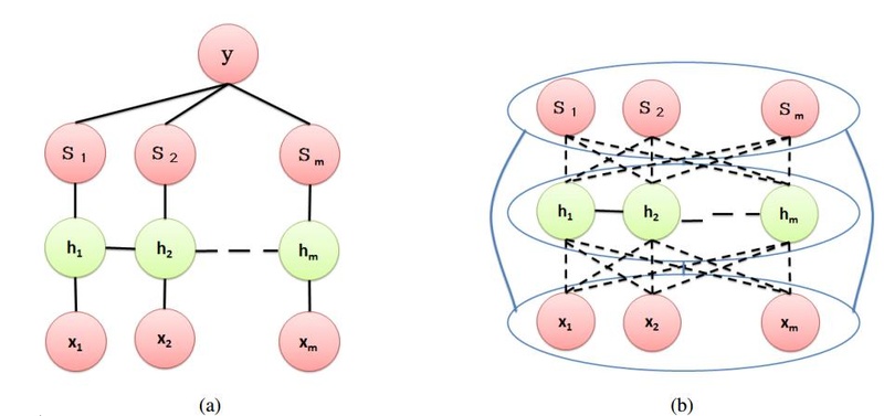
图a为CRF算法,图b为本文使用的算法,由第一层算法得到中间值hi,再参照原始信息通过第二层算法得到所要的分割结果,因此在精确性上得到了极大的提高。
演示结果:
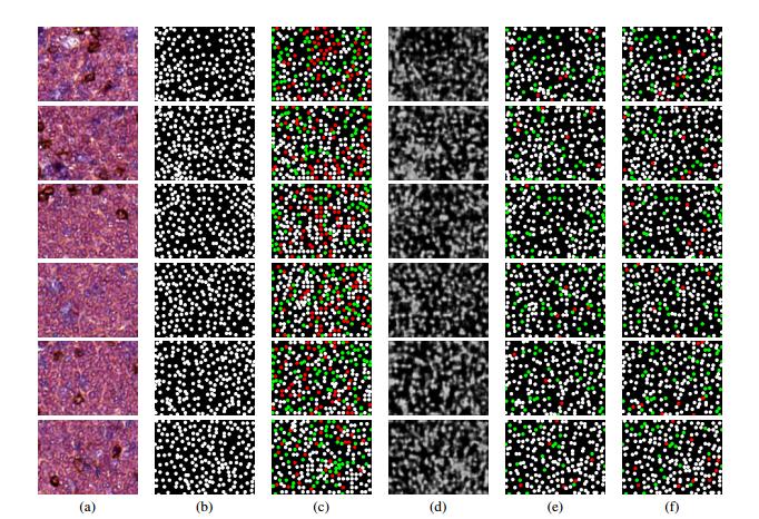
实验结果比较:(a)原始数据(b)标答(c)基于 Object Detection+Layout 算法的结果(d) 基于 Auto-context 算法的结果(e)基于双层模型(第二层算法使用置信度传播)算法的结果(f) 基于双层模型(第二层算法使用跳跃-扩散模型)的结果。其中圆点为细胞中心,红色点为错误识别的结果,绿色的点为漏识别的细胞中心,白色的点为正确的识别结果,从上图可见本算法具有极高的优越性,解决了细胞中心点分割的棘手问题。
A two-layer structure prediction framework for microscopy cell detection
Yan Xua, b,Weiying Wua,Eric I-Chao Changb, Danny Chenc,Jian Muc,Peter P. Leed,Kim R.M.Blenmand,Zhuowen Tue,f,*
a State Key Laboratory of Software Development Environment, Key Laboratory of Biomechanics and Mechanobiology of Ministry of Education, Beihang University, China
b Microsoft Research, Beijing, China
c Department of Computer Science and Engineering, University of Notre Dame, Notre Dame, USA
d Cancer Immunotherapeutics and Tumor Immunology (CITI), Duarte, CA, USA
e Department of Cognitive Science, University of California, San Diego, CA, USA
f Department of Computer and Engineering, University of California, San Diego, CA, USA
Abstract
The task of microscopy cell detection is of great biological and clinical importance. However, existing algorithms for microscopy cell detection usually ignore the large variations of cells and only focus on the shape feature/descriptor design. Here we propose a new two-layer model for cell centre detection by a two-layer structure prediction framework, which is respectively built on classification for the cell centres implicitly using rich appearances and contextual information and explicit structural information for the cells. Experimental results demonstrate the efficiency and effectiveness of the proposed method over competing state-of-the-art methods, providing a viable alternative for microscopy cell detection.
Keywords: Microscopy cell detection; Layered models; Structural prediction; Computer vision; Machine learning
 搜索
 导航